
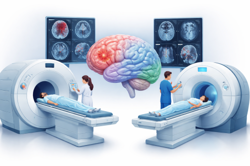
Medical imaging plays a crucial role in modern neurology. When patients experience symptoms such as headaches, dizziness, seizures, memory problems, or weakness, neurologists often recommend imaging tests to examine the brain. Two of the most common diagnostic tools are the CT scan (Computed Tomography) and MRI (Magnetic Resonance Imaging).
Although both tests create images of the brain and help doctors diagnose neurological conditions, they use very different technologies and are used in different clinical situations. Understanding the difference between an MRI and a CT scan can help patients feel more informed and less anxious when these tests are recommended.
What is a CT Scan?
A CT scan, also known as Computed Tomography, is a medical imaging test that uses X-rays and computer processing to create detailed cross-sectional images of the brain.
During the examination, the scanner rotates around the patient's head and captures multiple X-ray images from different angles. These images are then processed by a computer to produce slices of the brain that doctors can analyze.
One of the main advantages of a CT scan is its speed. The entire procedure usually takes only a few minutes, which makes it extremely useful in emergency situations.
Neurologists commonly use CT scans when they need to quickly detect:
- Brain bleeding
- Skull fractures
- Large brain tumors
- Stroke in the acute phase
- Head trauma
Because CT scans are fast and widely available in hospitals and emergency departments, they are often the first imaging test performed when a patient arrives with sudden neurological symptoms.
However, CT scans use ionizing radiation, which means they expose the body to a small dose of X-rays. While this exposure is generally considered safe when medically necessary, doctors avoid repeated CT scans unless they are clearly needed.
What is an MRI?
Magnetic Resonance Imaging (MRI) is another powerful imaging method used extensively in neurology. Unlike CT scans, MRI does not use radiation. Instead, it uses strong magnetic fields and radio waves to generate detailed images of the brain and spinal cord.
MRI works by detecting how hydrogen atoms in the body's tissues respond to magnetic energy. A computer then converts these signals into highly detailed images of the brain.
The main advantage of MRI is its exceptional ability to visualize soft tissues. This allows neurologists to detect subtle abnormalities that may not be visible on a CT scan.
MRI is often the preferred imaging test when doctors are investigating conditions such as:
- Multiple sclerosis
- Brain tumors
- Epilepsy
- Chronic headaches and migraines
- Neurodegenerative diseases such as Alzheimer’s disease
- Small strokes or microvascular brain changes
- Spinal cord disorders
Because MRI produces extremely detailed images of brain tissue, it is particularly valuable for diagnosing complex neurological conditions.
However, MRI scans take longer to perform than CT scans. The examination usually lasts between 20 and 45 minutes, and the patient must remain still during the procedure. In addition, MRI cannot be performed in patients who have certain types of metal implants, pacemakers, or other electronic devices.
When do Neurologists choose CT or MRI?
The choice between CT and MRI depends on the patient's symptoms, medical history, and the urgency of the situation.
In emergency cases such as head trauma, suspected brain bleeding, or acute stroke, doctors usually perform a CT scan first because it is fast and immediately available.
For more detailed evaluation of brain tissue or when investigating chronic neurological symptoms, neurologists often recommend an MRI scan because it provides much clearer images of the brain's structures.
In many cases, the two tests complement each other, and both may be used during the diagnostic process.
Are MRI and CT scans safe?
Both imaging methods are considered safe and routinely used worldwide.
CT scans involve a small amount of radiation, but the diagnostic benefits usually outweigh the risks when the test is medically necessary.
MRI does not expose patients to radiation, which makes it particularly useful when repeated imaging is required. However, patients must inform their doctor if they have metal implants, artificial joints, pacemakers, or other devices, as these may interfere with the magnetic field.
Why Brain imaging is important in Neurology
Brain imaging has revolutionized the field of neurology. These technologies allow doctors to examine the brain in detail and identify abnormalities that would otherwise remain hidden.
Imaging tests help neurologists:
- Detect structural brain abnormalities
- Identify vascular problems such as stroke
- Diagnose tumors or inflammation
- Evaluate neurological symptoms such as seizures or memory loss
- Monitor the progression of neurological diseases
Accurate imaging allows doctors to make faster diagnoses and start the most appropriate treatment as early as possible.
Both CT scans and MRI scans are essential diagnostic tools in neurology, but they serve different purposes.
CT scans are typically used for rapid evaluation in emergency situations, while MRI scans provide more detailed images that help diagnose complex neurological conditions.
If your neurologist recommends brain imaging, it is because these tests provide valuable information that helps guide diagnosis and treatment. Early and accurate imaging plays a key role in protecting brain health and improving patient outcomes.